
52 y/o male presents stating that he has had an area of redness on his right thigh that has “fever” in it. No systemic fever/SOB/CP. On physical exam, vitals are normal and there is a 5 cm patch of erythema and induration on the patient’s right anterior thigh. No lower extremity edema, no fluctuance of the area. You recall an article that showed 40% of patients with clinically suspected cellulitis were diagnosed with abscess via ultrasound and required incision and drainage, so you obtain the following scan. What’s the diagnosis?
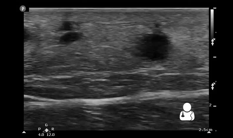

Diagnosis: Superficial Thrombophlebitis (STP)
This scan demonstrates a contiguous hypoechoic serpiginous structure, most consistent with dilated superficial veins. These veins appear to be noncompressible and are filled with heterogenously echogenic material, most likely thrombus. Further scans on this patient revealed no evidence of DVT.
- In cases of possible cellulitis or abscess, STP should be kept in the differential diagnosis.
- STP can be readily identified on bedside ultrasound, the most accurate finding is a noncompressible superficial vein.1
- Lower extremity STP has been shown to be associated with an increased risk of concomitant DVT (OR 3.3),2 particularly when it is above the knee,3 so further scanning via 2-3 point compression should be performed to rule out DVT in these patients. Furthermore, these patients should follow up to ensure resolution and rule out extension of disease to the deep veins.
- Barring any contraindication, prophylactic dose fondaparinux given for six weeks is a valid therapeutic option for STP of the legs. It “was associated with a significant reduction in symptomatic VTE (RR 0.15; 95% CI 0.04 to 0.50), extension (RR 0.08; 95% CI 0.03 to 0.22), and recurrence of ST (RR 0.21; 95% CI 0.08 to 0.54) with comparable rates of major bleeding.4“
- Lutter KS, Kerr TM, Roedersheimer LR, Lohr JM, Sampson MG, Cranley JJ. Superficial thrombophlebitis diagnosed by duplex scanning. Surgery. 1991;110:(1)42-6. [pubmed]
- Quéré I, Leizorovicz A, Galanaud JP, et al. Superficial venous thrombosis and compression ultrasound imaging. J Vasc Surg. 2012;56:(4)1032-8.e1. [pubmed]
- Bergqvist D, Jaroszewski H. Deep vein thrombosis in patients with superficial thrombophlebitis of the leg. Br Med J (Clin Res Ed). 1986;292:(6521)658-9. [pubmed]
- Di Nisio M, Wichers IM, Middeldorp S. Treatment for superficial thrombophlebitis of the leg. Cochrane Database Syst Rev. 2012;3:CD004982. [pubmed]
