
This patient presents with a severely depressed mental status and hypotension. The following ultrasound was obtained after an effective treatment was initiated. What was the treatment?

Answer: Transcutaneous Cardiac Pacing
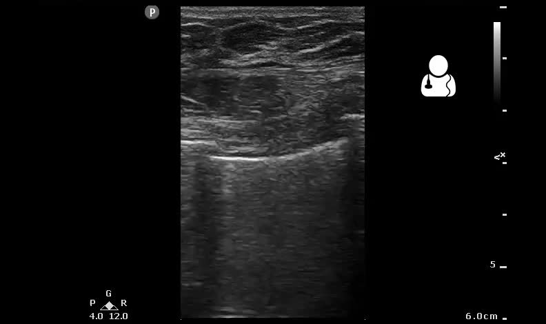
This left thoracic scan was performed using a linear transducer in a lung preset. There is obvious lung sliding, ruling out a pneumothrorax at this location. The pectoralis muscle is noted to twitch rhythmically as each transcutaneous electrical pulse is imparted to the chest wall.
- This is a great example of how much muscle twitching can be seen with transcutaneous pacing, demonstrating how easy it can be to mistake this twitching for a pulse. Ultrasound should be used to confirm electro-mechanical capture while pacing a critical patient. See also: UOTW #15.
- Small changes in the patient’s physiologic status (hypoxia, acidosis) can result in a heart that becomes recalcitrant to pacing, so these patients should still be considered critical and should be monitored extremely closely.1
- Note that this scan differs from a lung pulse, which is the rhythmic movement of the normally apposed visceral and parietal pleura as cardiac motion is transmitted through the lung. This sign can also can be used to rule out pneumothorax at that particular rib space. Unilateral lung pulse without lung sliding after endotracheal intubation suggests a main-stem intubation.2
